


Cancer battles look to high tech
- October 9, 2015
By Laureen Diephof | From The Salinas Californian
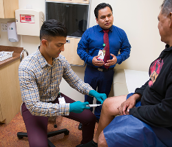
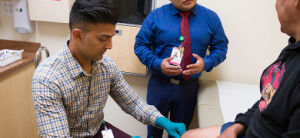
SALINAS, CA – October 9, 2015 – Because of advanced diagnostic technologies resulting in accurate and early detection, significant positive outcomes are coming to women diagnosed with breast cancer today in Monterey County. ● “The five-year survival rate for breast cancer for women who were diagnosed or treated is 89 percent,” said Amy Lantis Stemerman, M.D. of the Nancy Ausonio Mammography Center of Salinas Valley Memorial Health Care System in Salinas. ● Other Monterey County physicians, radiologists, nurses, sonographers and mammography technologists, share this good news. ● Advances in diagnostics include ultrasound, digital mammography, magnetic resonance imaging and several additional technologies new to our community.
Early Diagnosis
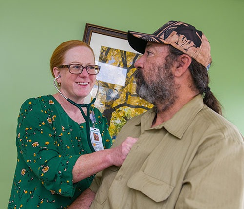
Let us imagine what early diagnosis looks like. A woman will walk into the Nancy Ausonio Mammography Center of SVMH in Salinas. Her general physician or another physician has referred her to the Center.
In a spa like setting that creates a calming, soothing ambiance, she relaxes before her name is called for the mammogram.
Additionally, while filling out the necessary paper work, a woman who identifies herself as having a family history of cancer has the option of completing a breast health screening form.
This form is forwarded to Bernadette A. Lucas Burch R.N. BSN. CBCN, the navigator who performs lifetime risk stratification, utilizing the TYRER-Cuzick risk calculator.
“The resulting score places the woman into an average, moderate or high lifetime risk category onto a template,” said Lucas Burch.
“A letter is then sent to the patient and their provider informing them of the findings. The radiologists’ recommendations for screening are given at that time as well,” added Burch.
A digital mammography ensures clear images. The scans and results can be viewed by physicians, securely online, from any computer, at any time.
“In many cases, breast cancer can be ruled out with a diagnostic mammogram, a breast ultrasound or breast MRI. If cancer has been ruled out, then the patient will wait for another year to get another mammogram. However, if cancer cannot be ruled out, the woman will need to have a biopsy,” said Dr. Amy Lantis Stemerman, M.D., medical director of the Nancy Ausonio Mammography Center, in Salinas.
According to Dr. Stemerman, a biopsy involves removing cells or tissue from the suspicious area of the breast. The cells or tissue are studied under a microscope to see if they show cancer.
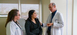
When cancer is diagnosed, the radiology, medical and radiation oncology, pathology and surgery meet in the conference room to discuss individual breast cancer cases. They review the person’s condition from every angle to determine the most effective treatment options.
“The goal of our breast health program is to empower women with personalized information that will assist them in making the best decisions,” said Dr. Stemerman.
“Target therapy” is the term used by medical director of the Carol Hatton Breast Care Center of the Community Hospital of the Monterey Peninsula (CHOMP) Dr. Grant Swanson.
Dr. Swanson uses this term in respect to the types of therapy used to match the exact treatment to the appropriate condition of the woman needing treatment.
“There are wide ranges of breast cancer,” according to Dr. Swanson, “and they may be subdivided into quite a few different types.”
With several types of treatments to help a woman diagnosed with breast cancer, whether it is a fast or slow growing type, it may or may not respond to treatments, such as hormonal treatment, for example. And then it may be that the woman needs chemotherapy, or another treatment.
“So there are critical questions to ask, and for that, a focus is placed on what treatment will match that woman’s cancer disease.”
According to Dr. Swanson, the new test, called Oncotype DX was recently published.
“This test examines a breast cancer patient’s tumor tissue at a molecular level.”
It helps physicians and patients decide on the best treatment for that particular women’s cancer, and the likelihood of recurrence.
Another new finding is the “HER2 cancer. This is one type of cancer that gives understanding about what makes an abnormality,” Dr. Swanson said.
HER2 is a cancer found in 10 to 15 percent of women, according to Dr. Swanson. This type of cancer tests for a protein called human epidermal growth factor, which promotes the growth of cancer cells.
“The knowledge of this cancer changed most untreatable to most treatable. The treatment of HER2 will change things around and dramatically prolong a woman’s life.”
Because of new treatment options, Dr. Swanson said, physicians are able to understand the inner workings of the cancer and what drives the behavior of a cancer cell.
“If we can see what is driving the cancer we are then given the opportunity to target that woman’s specific cancer cells.”
Survival odds improve with early screening
Early screening is most important, all experts agree, and with that, a recent law was passed concerning a woman who has dense breasts.
It is more difficult to see dense breasts from a mammogram, so the law instructs that doctors must notify the woman that they have dense breasts. When this is determined, the whole breast can be viewed with an ultra sound.
Ultrasound can locate the position of a tumor, which will guide the physician for biopsy.
If the cancer is removed surgically, is that the end of the story?
“After all is done surgically for the woman, it’s possible that the cancer had already spread, but so small we could not detect it. For some women, after surgery, we will treat for the risk of previously spread cancer,” Dr. Swanson explained.
“If we know what is broken, we can direct the treatment,” Dr. Swanson added.
A yearly mammogram may clear a woman of breast cancer or obtain an early diagnosis at the comfortable Carol Hatton Breast Care Center.
The Hologic Selenia Dimensions with AWS (Acquisition Work Station) 8000 system.
Natividad Medical Center has increased its ability to detect breast cancer in women at an early stage.
According to a press release, the new 3D digital is the only mammography system in Monterey County. It was in place and ready for October Breast Cancer Awareness Month.
The 3D mammography or breast tomosynthesis was installed Aug. 21 in the Radiology Department at Natividad Medical Center. Approved as an imaging tool by the Federal Drug Administration in 2011, the new 3D system, which is often used in combination with traditional 2D mammography, offers physicians a newer method to detect breast cancer at an early stage. According to the press release, a recent study of 25,000 women reported a 47% increase in cancer detection when tomosynthesis was used.
Digital tomosythesis helps to overcome the pressing of the breast during the mammogram. This has been unfavorable by some woman and might preclude some to get the annual test.
“We are beyond excited to get this advanced technology here at Natividad Medical Center,” said Heidi Riggenbach, Natividad Radiology Manager. “It not only gives us a more-efficient tool to detect cancer earlier, but it will give our patients peace of mind due to greater clarity and accuracy of the equipment. It will also decrease the number of callbacks which will spare women the anxiety, inconvenience and expense of coming back to the hospital for further imaging.”
Traditional 2D digital mammography takes images of the breasts in two dimensions and has been one of the most advanced tools in detecting breast abnormalities. In 3D mammography, the scanner moves in an arc over the breasts, taking multiple images from various angles. The images are displayed as a series of layers or thin slices, which can be viewed by radiologists one layer at a time, as a whole or in an interactive animation.
This improves the doctor’s ability to spot small abnormalities or tumors that are hidden by overlapping tissue and that may not be visible on standard mammograms. The greater sensitivity will also reduce false alarms and callbacks.
Studies in Europe and the U.S. have shown that combining 3D mammography with conventional 2D mammography can result in a 10 to 30% increase in overall breast cancer detection over 2D imaging alone, according to the press release.
Hologic, Inc., of Danbury, Connecticut, manufactures the Selenia Dimensions system. More than half of the U.S. News and World Report’s top 20 cancer hospitals offer Hologic 3D mammography systems. More than five million women to date in the U.S. have already been screened with this life-changing technology, which is now available in 50 states and in more than 50 countries.
Laureen Diephof is a freelance journalist living in Aromas. Contact her at ldiephof@sbcglobal.net.
Editor’s Note: This special series of articles focusing on Breast Cancer Awareness Month continues next Saturday with Edward Moncrief writing about the tragedy of breast cancer from the husband’s point of view.